Home
/ Pelvis Anterior View : Anatomy And Physiology I Bones Of The Pelvic, The pelvic bones are smaller and.
Pelvis Anterior View : Anatomy And Physiology I Bones Of The Pelvic, The pelvic bones are smaller and.
Pelvis Anterior View : Anatomy And Physiology I Bones Of The Pelvic, The pelvic bones are smaller and.. The hip bone, or coxal bone, forms the pelvic girdle portion of the pelvis. When hip flexors shorten, hip extensors loosen up or lengthen. Our latest youtube film is ready to run. The judet view is comprised of two projections, first the iliac oblique for assessment of the posterior column and anterior wall of the acetabulum; The pelvis is a ring structure made up of three bones:
In this image, the pelvis is shown as it would be in the erect posture. The hip bone, or coxal bone, forms the pelvic girdle portion of the pelvis. Illustration of left hemipelvis and hip joint, anterior view, showing bones: This stock medical illustration shows an anterior (front) view of the female abdomen and pelvis. The prominent bones and bony landmarks have been labeled.
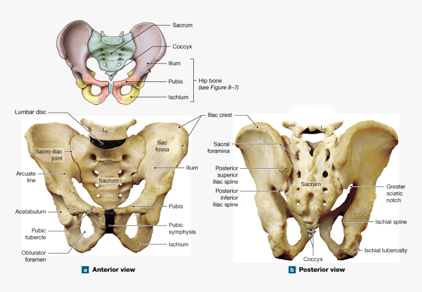
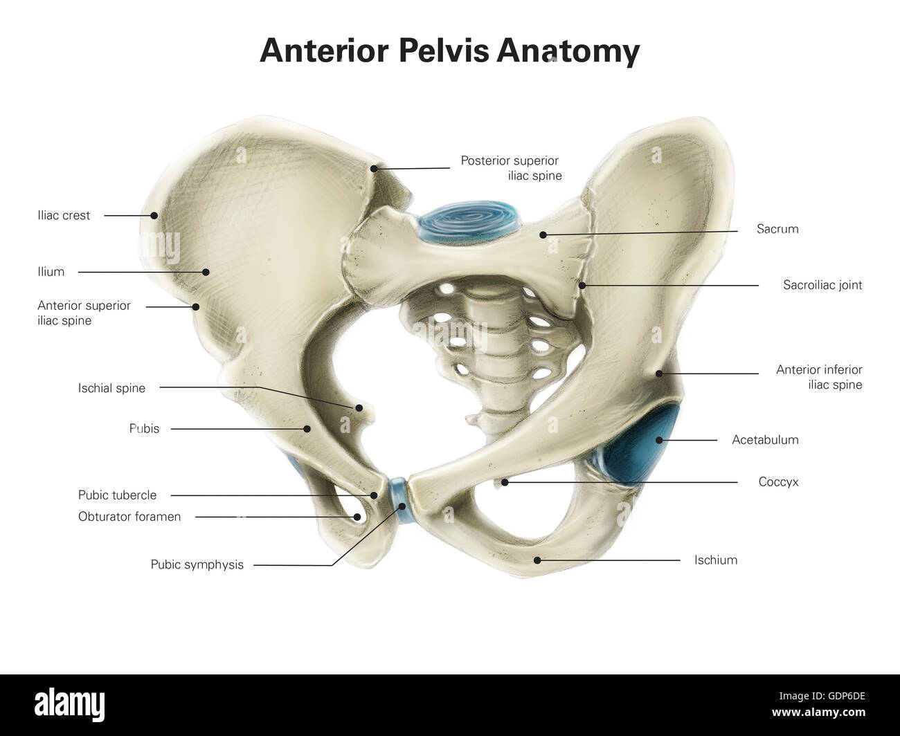
Figure 8 6 The Pelvis Anterior View Hd Png Download Kindpng from www.kindpng.com The anterior superior iliac spine and pubic tubercle are in the same vertical plane. Together, they form the part of the pelvis called the pelvic girdle. The hip bone, or coxal bone, forms the pelvic girdle portion of the pelvis. Superior ramus of pubis, pubic crest, inferior ramus of pubis, ischial ramus, obturator foramen, ischial tuberosity, iliac crest, iliac tubercle, anterior superior iliac spine, posterior inferior iliac spine should be iliac spine?, femoral head, greater trochanter, neck, lesser. Browse 4,822 hip anatomy stock photos and images available, or search for hip replacement or knee anatomy to find more great stock photos and pictures. The ap pelvis view is part of a pelvic series examining the iliac crest, sacrum, proximal femur, pubis, ischium and the great pelvic ring. The pelvic region is the area between the trunk — or main body — and the lower extremities, or legs. In this image, you will find the posterior superior iliac spine, iliac crest, tubercle of the iliac crest, anterior superior iliac spine, greater sciatic foramen, the acetabular margin in it.
View of the pelvic outlet and pelvic muscles from below.
This stock medical illustration shows an anterior (front) view of the female abdomen and pelvis. Human hip and pelvis anterior view of the hip and pelvis, showing attachment of ligaments to the femur, ilium, ischium, and pubis. In this image, the pelvis is shown as it would be in the erect posture. View of the pelvic outlet and pelvic muscles from below. The pelvic skeleton is formed in the area of the back, by the sacrum and the coccyx and anteriorly and to the left and right sides, by a pair of hip bones. The anterior superior iliac spine and pubic tubercle are in the same vertical plane. It allows for complete rotations of the hip and is also. The prominent bones and bony landmarks have been labeled. The posterior tibial allows the foot to extend. Looking at the pelvis from the inside, you should be able to identify the following items: Learn vocabulary, terms, and more with flashcards, games, and other study tools. When a muscle tightens, it shortens. The paired hip bones are the large, curved bones that form the lateral and anterior aspects of the pelvis.
Anterior tibialis thick muscle enabling the foot to flex on the leg and to draw near the median axis of the body; This is a free printable worksheet in pdf format and holds a printable version of the quiz ligaments of the pelvis (anterior view).by printing out this quiz and taking it with pen and paper creates for a good variation to only playing it online. The bones of the pelvis and lower back work together to support the body's weight, anchor the abdominal and hip muscles, and protect the delicate vital organs of the vertebral and abdominopelvic cavities. The sacroiliac joints are true synovial joints, but the symphysis is a synchrondrosis, without a synovial space. Learn vocabulary, terms, and more with flashcards, games, and other study tools.
Pelvis Skeleton Anterior View from www.anatomynote.com This is an online quiz called ligaments of the pelvis (anterior view) there is a printable worksheet available for download here so you can take the quiz with pen and paper. Human hip and pelvis anterior view of the hip and pelvis, showing attachment of ligaments to the femur, ilium, ischium, and pubis. The vertebral column of the lower back includes the five lumbar vertebrae, the sacrum, and the coccyx. This is a free printable worksheet in pdf format and holds a printable version of the quiz ligaments of the pelvis (anterior view).by printing out this quiz and taking it with pen and paper creates for a good variation to only playing it online. The anterior superior iliac spine and pubic tubercle are in the same vertical plane. The pelvis is a ring structure made up of three bones: The pelvic region is the area between the trunk — or main body — and the lower extremities, or legs. The judet view is comprised of two projections, first the iliac oblique for assessment of the posterior column and anterior wall of the acetabulum;
The pelvis is a ring structure made up of three bones:
The ap pelvis view is part of a pelvic series examining the iliac crest, sacrum, proximal femur, pubis, ischium and the great pelvic ring. Bony pelvis or pelvic skeleton is formed by hip bones, sacrum and coccyx. The posterior tibial allows the foot to extend. Human hip and pelvis anterior view of the hip and pelvis, showing attachment of ligaments to the femur, ilium, ischium, and pubis. In this image, you will find the posterior superior iliac spine, iliac crest, tubercle of the iliac crest, anterior superior iliac spine, greater sciatic foramen, the acetabular margin in it. This stock medical illustration shows an anterior (front) view of the female abdomen and pelvis. Secondly, the obturator oblique view demonstrating the anterior column of the pelvis along with the posterior wall of the acetabulum. Aiis, anterior inferior iliac spine; The anterior superior iliac spine and pubic tubercle are in the same vertical plane. Sa110004 fotosearch stock photography and stock footage helps you find the perfect photo or footage, fast! The pelvic region is the area between the trunk — or main body — and the lower extremities, or legs. The judet view is comprised of two projections, first the iliac oblique for assessment of the posterior column and anterior wall of the acetabulum; Learn vocabulary, terms, and more with flashcards, games, and other study tools.
The pelvis is a ring structure made up of three bones: The judet view is comprised of two projections, first the iliac oblique for assessment of the posterior column and anterior wall of the acetabulum; The hip bone, or coxal bone, forms the pelvic girdle portion of the pelvis. Together, they form the part of the pelvis called the pelvic girdle. Anterior view of the lumbar spine and pelvis.
Anterior View Of Human Pelvis With Labels Stock Photo Alamy from c8.alamy.com The pelvis has three joints: The pelvis is a ring structure made up of three bones: The pelvic region is the area between the trunk — or main body — and the lower extremities, or legs. Illustration about anatomy, bone, posterior, anterior, foramen, ischium, pelvic, pelvis, ilium, health, sacro, lumbosacral, human. This is an online quiz called ligaments of the pelvis (anterior view) there is a printable worksheet available for download here so you can take the quiz with pen and paper. This stock medical illustration shows an anterior (front) view of the female abdomen and pelvis. The sacroiliac joints are true synovial joints, but the symphysis is a synchrondrosis, without a synovial space. Secondly, the obturator oblique view demonstrating the anterior column of the pelvis along with the posterior wall of the acetabulum.
When hip flexors shorten, hip extensors loosen up or lengthen.
Anterior view of a woman's pelvis. Learn vocabulary, terms, and more with flashcards, games, and other study tools. This is a free printable worksheet in pdf format and holds a printable version of the quiz ligaments of the pelvis (anterior view).by printing out this quiz and taking it with pen and paper creates for a good variation to only playing it online. Our latest youtube film is ready to run. Superior ramus of pubis, pubic crest, inferior ramus of pubis, ischial ramus, obturator foramen, ischial tuberosity, iliac crest, iliac tubercle, anterior superior iliac spine, posterior inferior iliac spine should be iliac spine?, femoral head, greater trochanter, neck, lesser. The pelvis has three joints: The anterior superior iliac spine and pubic tubercle are in the same vertical plane. This stock medical illustration shows an anterior (front) view of the female abdomen and pelvis. Anterior tibialis thick muscle enabling the foot to flex on the leg and to draw near the median axis of the body; Two sacroiliac joints and the pubic symphysis. The sacroiliac joints are true synovial joints, but the symphysis is a synchrondrosis, without a synovial space. There is a triangular sheet of dense musculofascial tissue that spans the anterior half of the. When a muscle tightens, it shortens.
